Our work

Neurons can be grouped by many features, such as transcriptional identity, physiology, and morphology. This includes their connectivity profile, which ultimately determines how neurons participate in a circuit. Our lab studies how synaptic connectivity is established during development, the specific molecular mechanisms that shape that process, and which of those mechanisms are conserved across species versus unique to humans. We're particularly interested in how these features of connectivity shape circuit function underlying learning and cognition, and how it may lead to neurodevelopmental disease vulnerability.

Human-specific genes and cortical connectivity

The human brain differs from other species through molecular changes that act at multiple stages of cortical development — from how neurons are generated to how they ultimately wire into circuits. We study several such human-specific genetic changes and how they shape brain development, function, and disease vulnerability.

Human-specific genes can shape how neurons wire together. A duplication unique to the human genome gave rise to SRGAP2C, a truncated, human-specific gene that interferes with its ancestral counterpart SRGAP2A. This human-specific paralog slows synaptic maturation and increases the density of both excitatory and inhibitory synapses on cortical pyramidal neurons, a change that, in mice carrying the human gene, leads to altered sensory processing and enhanced sensory-guided learning (Schmidt et al., Nature, 2021).

More recently, we've extended this work from single synapses to network-level activity. Using wide-field mesoscale imaging, we find that SRGAP2C reconfigures large-scale cortical network dynamics, changing how activity is distributed across cortical areas and how strongly different regions drive one another (Zhao, Anderson & Schmidt, bioRxiv, 2026).

Looking ahead, connectivity sits at the intersection of nearly every way we define a neuron's identity — its transcriptional profile, its synaptic proteome, its physiology are all shaped by who it connects to and how. We think this makes connectivity a particularly powerful lens for understanding what makes human cortical circuits distinctive. It's also where our understanding is most limited: we still know very little about which features of cortical connectivity are conserved across species and which are uniquely human. Going forward, we're working toward mapping cortical circuits at scale, combining transcriptomics, connectomics, and synaptic proteomics to build a more complete picture of circuit architecture, and asking how specific connectivity motifs make certain cell types and circuits selectively vulnerable in disease.
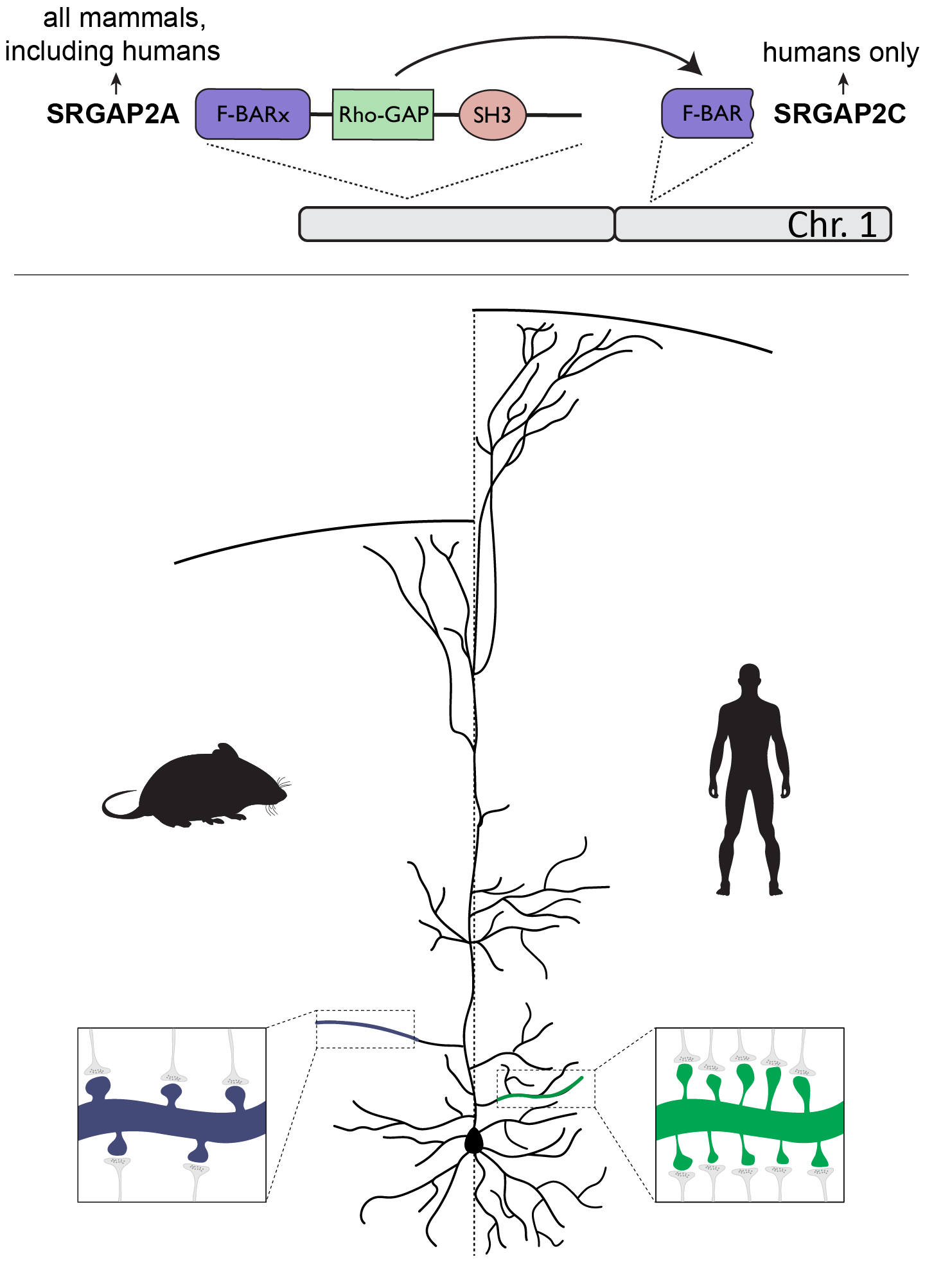
SRGAP2A to SRGAP2C human-specific gene duplication on chromosome 1

The ancestral copy SRGAP2A, which is located on chromosome 1, is present in most mammals. Duplication of SRGAP2A in the Homo lineage resulted in the emergence of multiple copies, one of which is SRGAP2C. Humans, as the only extant Homo species, are the only ones who possess this copy. Expression of SRGAP2C in mouse cortical pyramidal neurons leads to increased synaptic density and protracted synaptic maturation. This is akin to human cortical pyramidal neurons, which among other features, are characterized by a larger number of synapses that mature at a slower rate.
Molecular regulation of synapse-specific connectivity

Il1rapl2 is a synaptic protein with a strikingly specific expression pattern: unlike its broadly-expressed paralog Il1rapl1, Il1rapl2 is found almost exclusively in one class of deep-layer cortical neurons: the intratelencephalic (IT) neurons that carry the long-range, cortico-cortical projections thought to underlie higher-order brain integration. Both genes have been linked to autism spectrum disorder and intellectual disability, but the role of Il1rapl2 in shaping cortical circuits had not been directly tested.

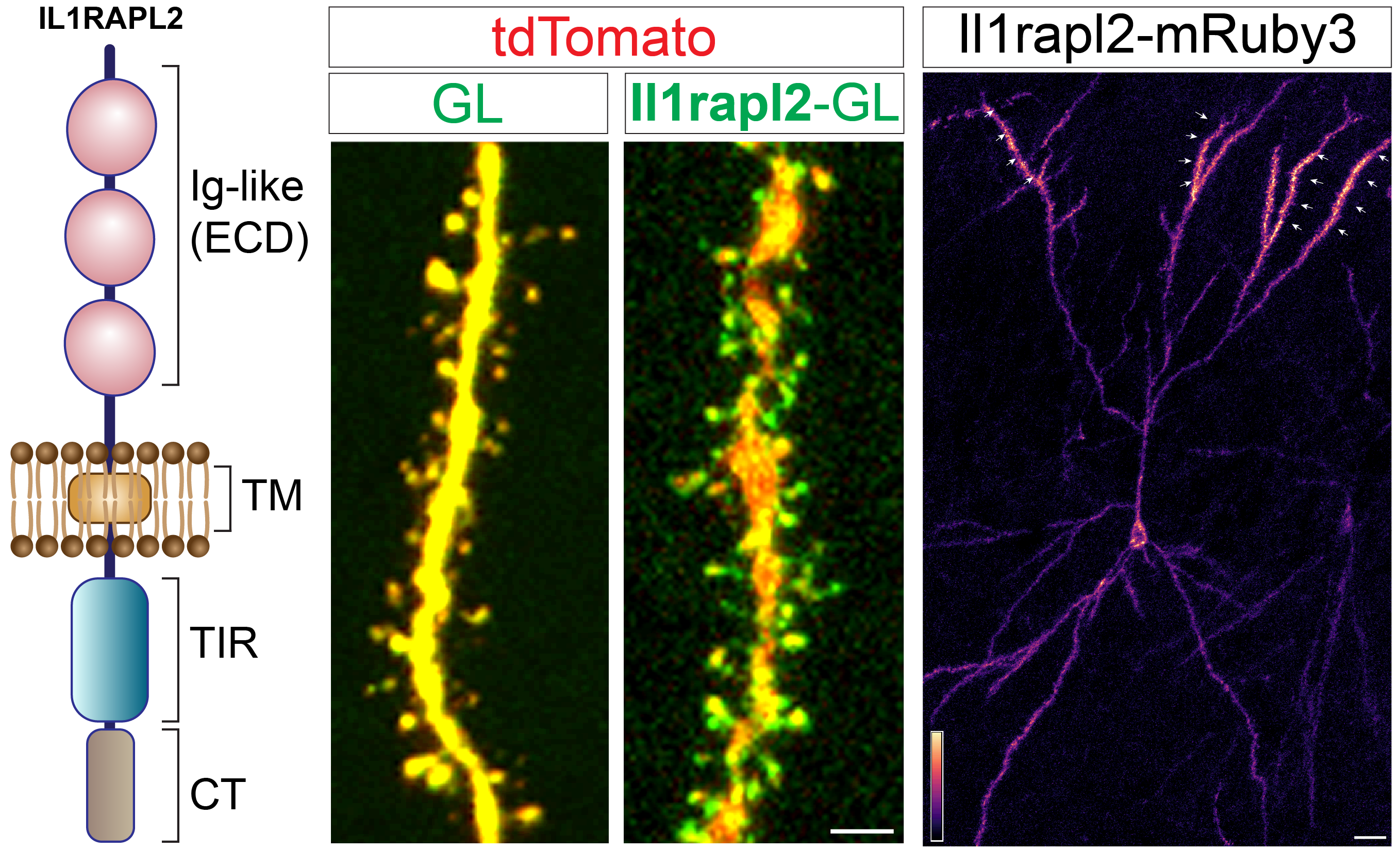
In collaboration with the Berto lab (MUSC), our early work shows that Il1rapl2 protein is concentrated specifically in the apical dendrites of IT neurons, where it localizes to dendritic spines, the sites of excitatory synapses.

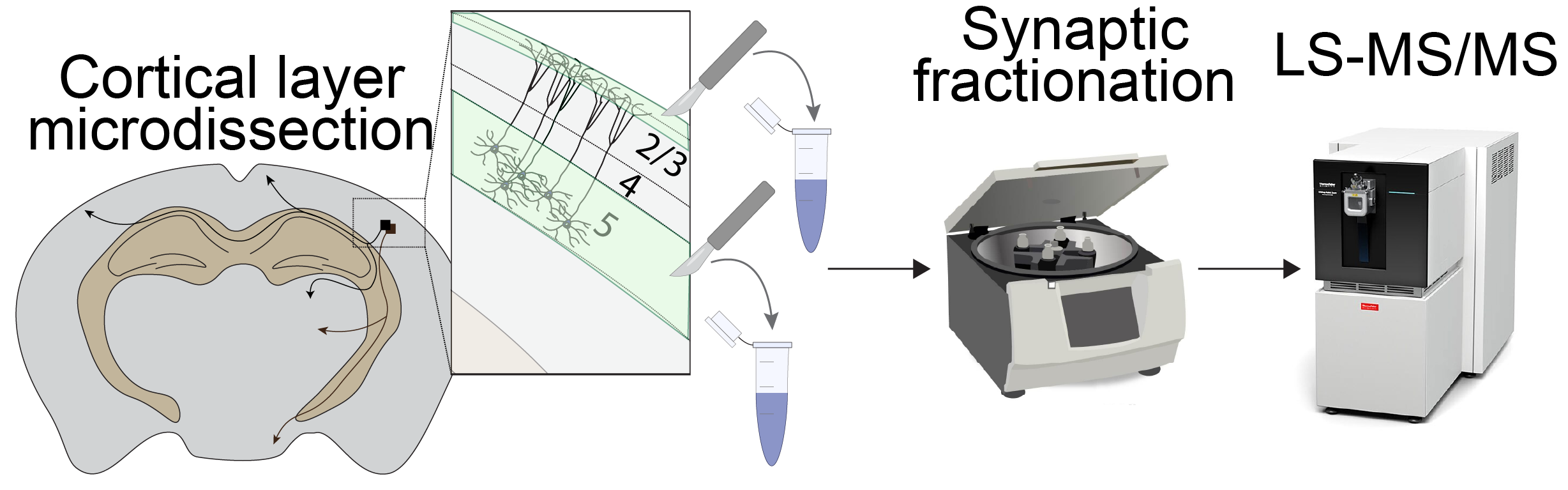
Molecular biology and proteomics are central to how we're pursuing this work. To determine what makes Il1rapl2-containing synapses molecularly distinct, we combine two complementary approaches: CRISPR-based tagging of the endogenous Il1rapl2 protein for direct visualization in neurons, and layer-specific microdissection paired with mass spectrometry to directly measure Il1rapl2 and its synaptic protein neighbors in isolated synaptic fractions. This lets us ask which other synaptic proteins are recruited alongside Il1rapl2 to build this specific class of long-range synapse, and how that molecular composition changes when Il1rapl2 is lost, directly linking specific molecular players at the synapse to the structural and functional specificity of connectivity.

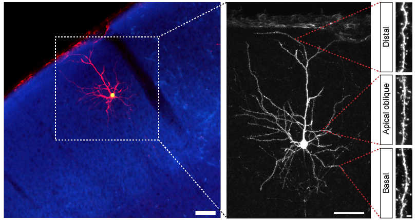
Il1rapl2-mRuby3 expression showing apical dendrite enrichment in a cortical IT neuron

Il1rapl2 is a postsynaptic transmembrane protein localized to dendritic spines of deep-layer cortical pyramidal neurons. Il1rapl2 tagged with mRuby3 shows strong enrichment of the protein in the apical dendritic domain.

How we study this

See below on how we use a combination of circuit mapping, in vivo imaging, and behavioral approaches to tackle these questions.

Two-photon microscopy

We use two-photon microscopy to investigate neuronal response properties at a cellular level.

two-photon imaging

Two-photon microscopy lets us image neuronal structure and activity in vivo with single-cell resolution, resolving individual neuron activity within the intact, living brain. This complements wide-field imaging (see below), which captures activity across the entire dorsal cortex.

Neuronal activity imaged in vivo in the barrel field of the somatosensory cortex.
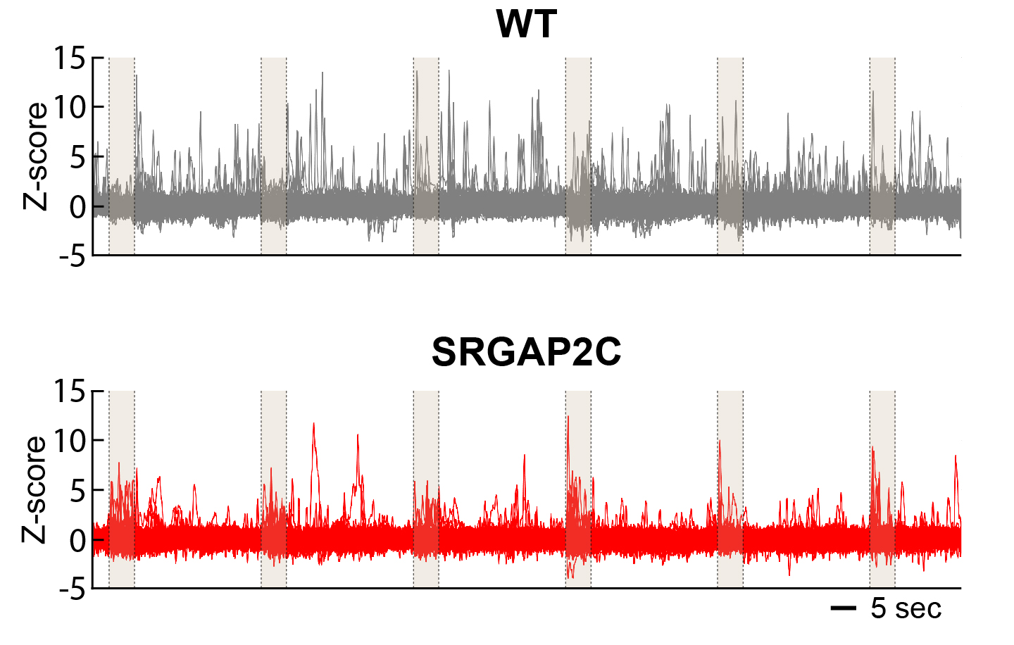
Our previous work showed that expression of the human-specific gene SRGAP2C increases sensory coding in layer 2/3 pyramidal neurons of the somatosensory cortex: SRGAP2C increases the neuronal response probability to whisker stimulation while reducing overall spontaneous activity.
neuronal response properties upon SRGAP2C expression

Expression of SRGAP2C increases the response probability to whisker stimulus (peaks in shaded areas) while reducing overall spontaneous activity (peaks outside shaded areas).
We're extending this approach to ask how the strength of specific long-range synaptic inputs onto individual dendritic compartments changes across development and with genetic manipulation.

Wide-field microscopy

Using wide-field microscopy we investigate how neuronal activity develops and propagates across the cortex.

wide-field optical mapping

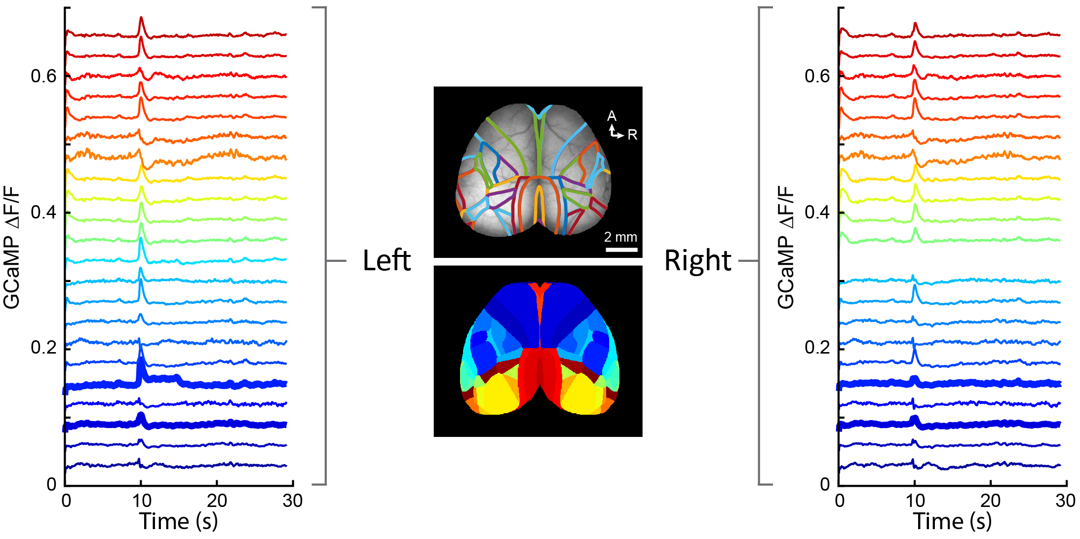
In order to examine functional connectivity across the cortex of mice, we use wide-field microscopy (WFM) to image neuronal activity in vivo across the entire dorsal cortex. WFM enables high-speed imaging over large fields of view and is therefore excellent for assessing spatiotemporal dynamics of neuronal activity across a large number of cortical regions. By parcellating the cortex into distinct brain regions we measure how neuronal activity emerges in each cortical region and how it subsequently spreads and propagates throughout the cortex.

Cortical activity patterns under resting-state conditions show rapid, spontaneous patterns of neuronal activity that are often bilaterally symmetrical.

parcellation of cortex

Parcellation of the dorsal cortex according to the Allen Brain Atlas.
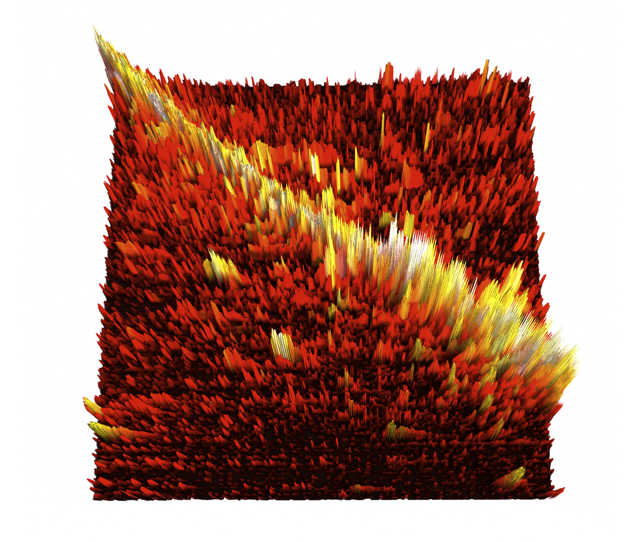
We've used this approach to show that expressing the human-specific gene SRGAP2C reorganizes how cortical regions communicate during behavior: task-related activity becomes more broadly distributed across cortical regions, and more strongly influenced by frontal areas, with sensory representations becoming more distinct as an animal transitions from sensing to acting (Zhao, Anderson & Schmidt, bioRxiv, 2026). We're extending this approach to ask how circuit-restricted regulators such as Il1rapl2 affect long-range functional coupling between cortical regions.

Neuronal circuit mapping

By employing a variety of tracing techniques we map structural connectivity throughout the brain.

Neuronal circuit mapping

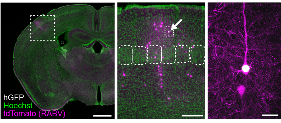
Using sparse in utero electroporation and viral tracing techniques, including whole-brain monosynaptic rabies tracing, we map connectivity across the brain at both a single-neuron and whole-brain level. Using this approach, we've shown that SRGAP2C selectively increases feedforward and feedback cortico-cortical connectivity, projections thought to be especially important for human cognition.
labeling of individual cortical neuron

Sparse in utero electroporation techniques let us visualize neuron morphology in high detail, up to individual synapses.
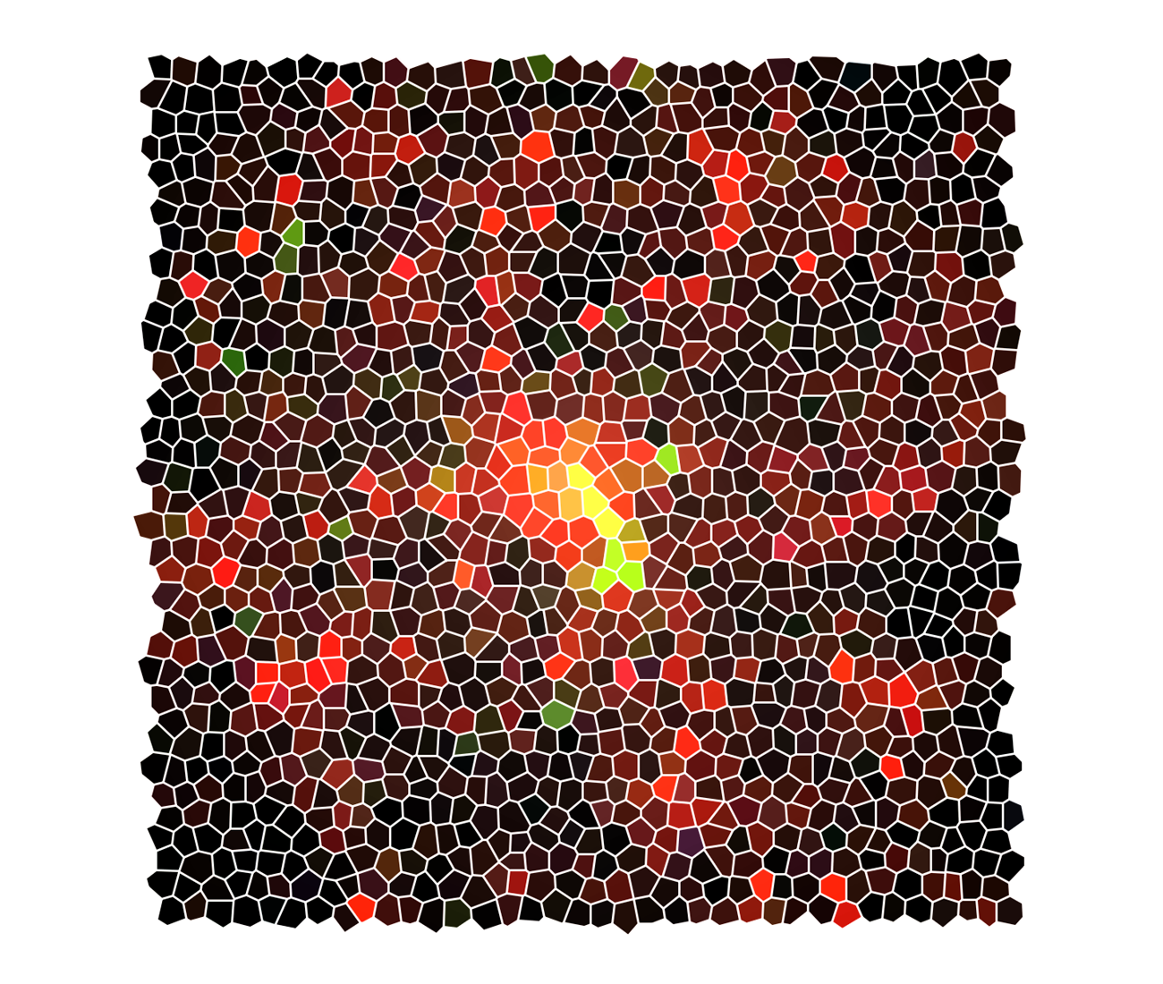
sparse monosynaptic rabies tracing

Monosynaptic rabies tracing maps the presynaptic connectivity (magenta) for a specific cortical neuron (hGFP) in mouse primary somatosensory cortex.

Whole-brain reconstruction registers every traced neuron onto the Allen Brain Atlas, letting us quantify how circuit organization changes across the brain.
We're now applying the same whole-brain tracing approach to Il1rapl2, to directly map which long-range inputs are lost when this synaptic regulator is missing.

Synaptic proteomics

We use mass spectrometry to directly measure the molecular composition of specific synapse types.

Synaptic proteomics workflow thumbnail

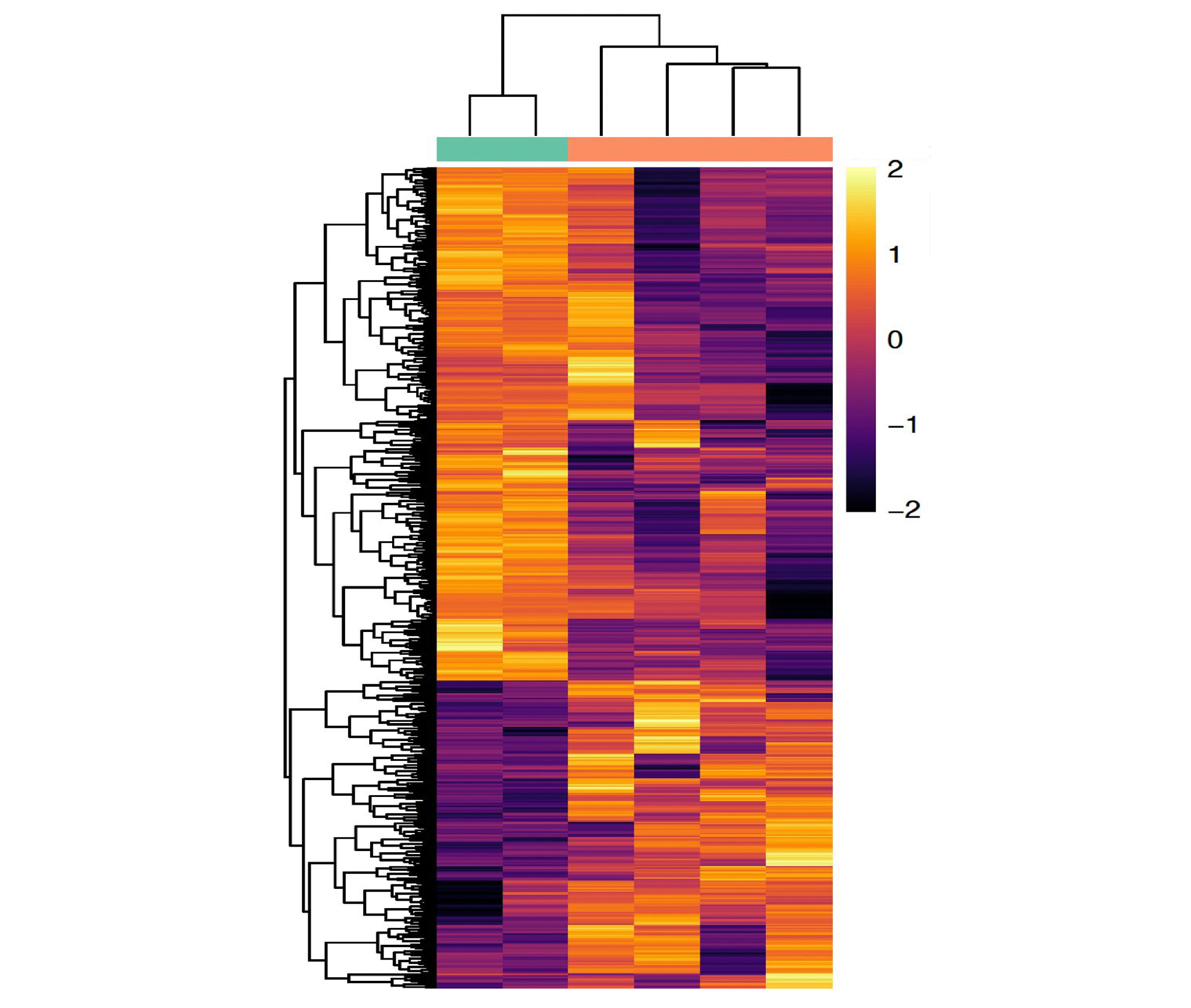
To understand what makes specific classes of synapses molecularly distinct, we combine synaptosomal fractionation — biochemically isolating synapses from specific cortical layers or dendritic compartments — with tandem mass spectrometry (LC-MS/MS) to directly identify and quantify the proteins present.
This approach lets us detect endogenous protein levels, compare synaptic protein composition across genotypes, and identify candidate molecular partners recruited to specific synapse types. We use it alongside CRISPR-based endogenous protein tagging and imaging to connect specific molecular players at the synapse to the structural and functional specificity of connectivity.
Synaptic proteomics workflow thumbnail

Behavior

Using a variety of behavioral paradigms we study how changes in neuronal circuit structure and function impact behavior.

mouse behavior

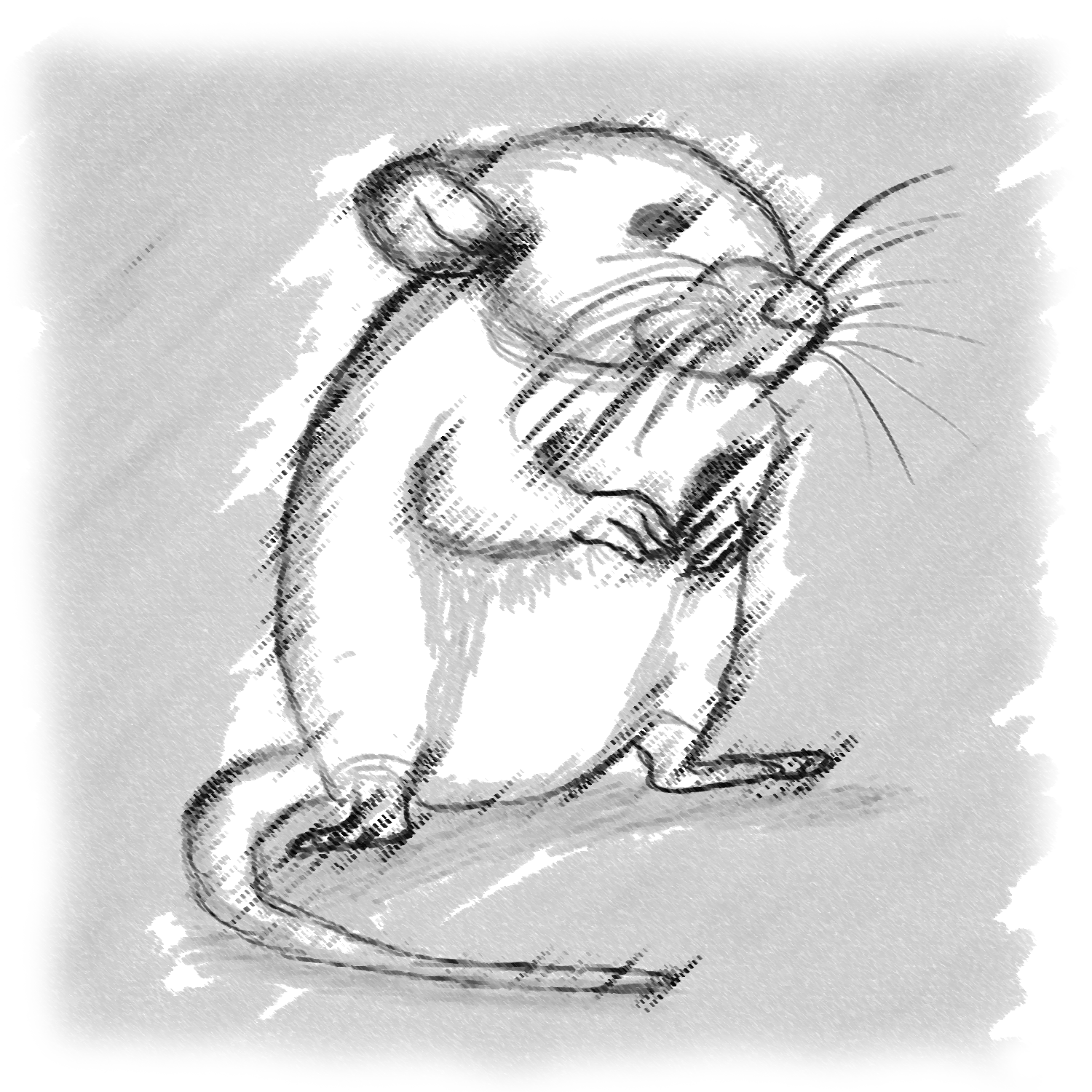
Behavior is how the brain's internal computations become visible: changes in circuit structure and function can directly alter what an animal is able to do, and how well.
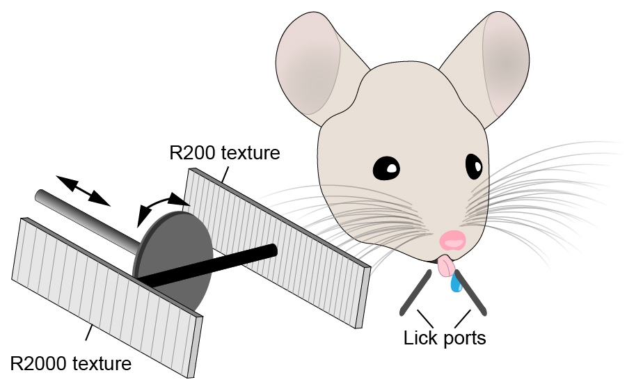
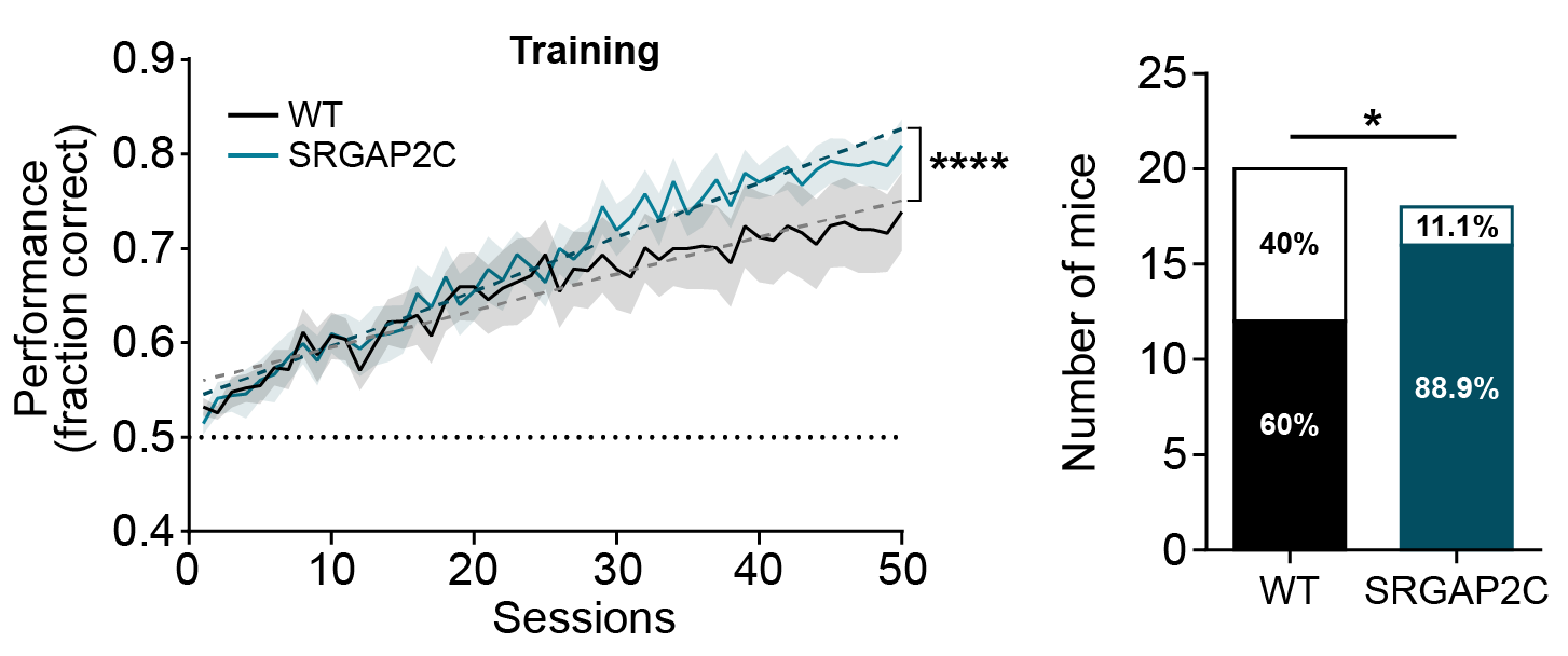
Our lab uses a variety of behavioral paradigms to study how human-specific genes that modify brain structure and function impact behavioral outcome — for example, showing that humanizing mice for SRGAP2C expression enhances performance in a difficult texture-discrimination task.
schematic of texture discrimination task

Mice are highly capable of discriminating textures with only their whiskers, in a task that can be made progressively harder.
behavioral performance of humanized SRGAP2C mice

While only 60% of wild-type mice learn this task over 50 sessions, almost all SRGAP2C mice are able to perform it.
Because impaired synaptic development is also a hallmark of neurodevelopmental disorders such as autism spectrum disorder, understanding how genetic changes affect behavior is central to understanding how such disorders arise.